Comparing EEG and MRI: Which is Right for Your Neurological Assessment?
Understanding EEG and MRI
When it comes to neurological assessments, two of the most common tools used are Electroencephalography (EEG) and Magnetic Resonance Imaging (MRI). Both are invaluable in diagnosing and monitoring neurological conditions, but they serve different purposes and provide distinct types of information. Understanding their differences can help in determining which is the right choice for your specific needs.
EEG and MRI are both non-invasive methods of examining brain function and structure. However, they operate on different principles. EEG records electrical activity in the brain, offering real-time data on how the brain responds to various stimuli. In contrast, MRI uses powerful magnets and radio waves to create detailed images of the brain's structure, providing insights into anatomical abnormalities.
Advantages of EEG
EEG is particularly effective for diagnosing conditions related to abnormal brain electrical activity, such as epilepsy. It is a relatively quick procedure and can be conducted at a lower cost compared to an MRI. The high temporal resolution of EEG allows for the detection of rapid changes in brain activity, making it useful for monitoring the brain's response to specific tasks or stimuli.
Another significant advantage of EEG is its ability to be performed in a variety of settings, including hospitals, clinics, and even at home in some cases. This flexibility makes EEG a versatile tool for ongoing monitoring of neurological conditions.

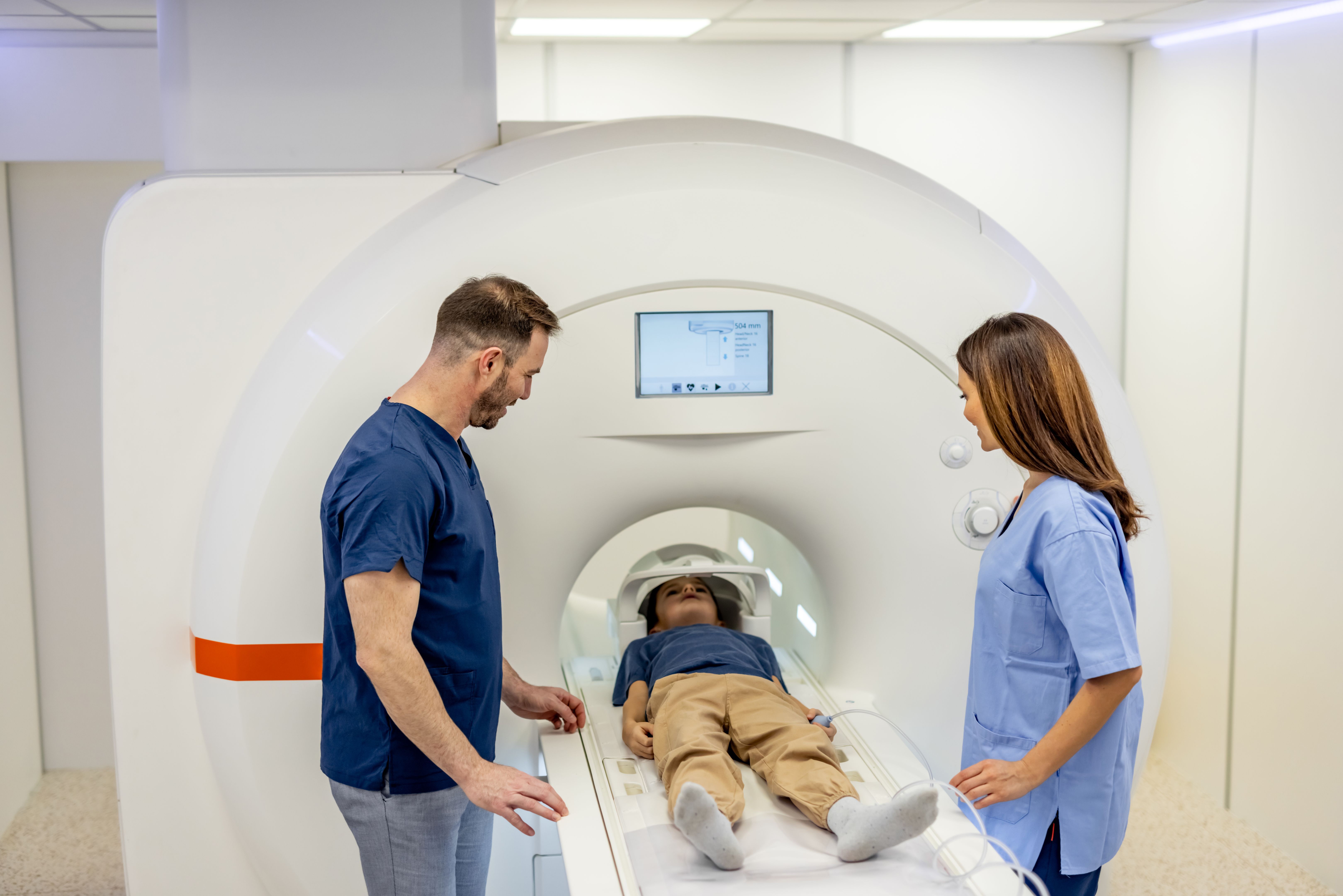
Benefits of MRI
MRI is renowned for its superior spatial resolution, which enables healthcare professionals to visualize the brain's structure in great detail. This capability makes MRI particularly useful for identifying tumors, brain injuries, and structural abnormalities. Unlike EEG, MRI does not involve exposure to radiation, making it a safer option for repeated use.
Moreover, advancements in MRI technology have led to functional MRI (fMRI), which allows for the observation of brain activity by measuring changes in blood flow. This technique provides a more comprehensive view of both brain structure and function, which can be crucial in complex cases.
Choosing the Right Assessment
The decision between EEG and MRI often depends on the specific clinical question at hand. If the primary concern is related to electrical activity or seizure disorders, an EEG may be more appropriate. Conversely, if structural abnormalities or detailed anatomical insights are required, an MRI would be the preferred choice.
In some cases, a combination of both EEG and MRI may be recommended to gain a more comprehensive understanding of a patient's neurological condition. Consulting with a neurologist can provide personalized guidance based on individual clinical needs.
Conclusion
Both EEG and MRI have their unique strengths and limitations. Understanding these differences is crucial for selecting the most appropriate method for neurological assessment. Whether it's the temporal precision of EEG or the detailed imagery provided by MRI, each tool offers valuable insights that contribute to accurate diagnosis and treatment planning.
Ultimately, the choice between EEG and MRI should be made in collaboration with medical professionals who can tailor their recommendations to your specific health needs. Staying informed about these diagnostic tools empowers patients to make educated decisions about their neurological care.